The End of All Disease |
m | ||||||||||||
Posted to Subscribers on 27 March 2008 |
|||||||||||||
|
|
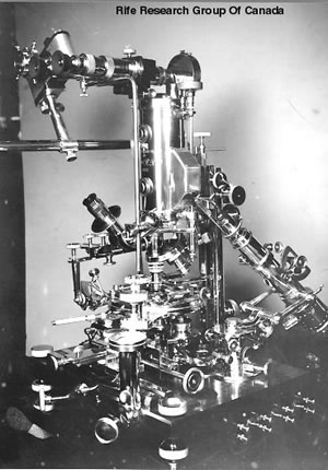
Seventy-seven years ago, forty-four of the country's most respected doctors and scientists attended a banquet at the estate of Dr. Milbank Johnson in Pasadena, California. The guest of honor was Dr. Royal Raymond Rife, and the occasion was the celebration of the end of all disease. A few years later, 1934, a committee from the University of Southern California brought 16 terminally ill cancer patients to Rife for treatment, with the intent of evaluating the success of his modalities. In the 90 days covered in the study, 14 patients, i.e., 86.5% of the patients, were deemed to have been completely cured. The treatment was then adjusted and the remaining two were cured during the next four weeks. By 1939, the prestigious scientists who had witnessed these "miracles" were denying that they had met Rife. How does a treatment this promising go underground where the risk of it being forgotten forever looms so large? Rife's Inventions Rife's life work consisted of two key inventions, both quite original. The one part was used for evaluation purposes. It was a very complex microscope that allowed users to view liquid samples without any fixatives. In short, the samples could be live, meaning a drop of blood could be viewed for hours or days and Rife spent countless hours every day making the sorts of detailed observations that his microscope rendered possible.
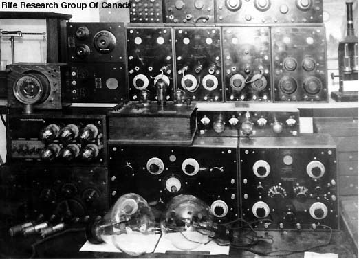
As early as 1920, Rife had identified a virus that he believed was the causative agent of cancer. He called it the BX virus. To give some idea of the perseverance and dedication of this scientist, Rife tried over and over and over again to force normal cells to become malignant. He tried 20,000 times and failed 20,000 times. However, when he irradiated the BX virus and transplanted it into mice, he succeeded 400 times in a row. The next task was to reverse or destroy the tumors. He postulated that everything has a unique vibratory rate and that there is a specific rate that is fatal for each particular organism. He called this the Mortal Oscillatory Rate. To create these rates, he had another invention, one consisting of a frequency generator and tubes containing noble gases.
In short, the microscope was used to understand what cannot be seen with the naked eye, but the treatment relied on the frequencies. The two pieces of equipment are not actually mutually dependent one upon the other and what is interesting is that today the microscopy is being carried on relatively independently of the vibratory treatments, mainly in Canada and Europe but to some extent in the U.S. despite ceaseless, senseless, and mindless persecution of those who dare to look where others have not. On the other hand, the frequency treatments have been much more resilient in the U.S. though still persecuted under the guise of protecting the public.
The era between the two world wars was full of foment. What is little realized is that the future of medicine was not, at that time, carved in stone. Vigorous debates over whether the cure for disease would be based on chemistry, aka pharmaceuticals, or electricity were occupying the minds of serious thinkers. Since the work of Pasteur and the Curies at the end of the 19th century, there was abundant attention on germs and mechanisms of destruction, but there was not at that time consensus around which strategies were most promising. Homeopathy was popular and the idea of energy was not actually outside the mind sets of thinking people, even in those unsettled times. Moreover, both chiropractic and osteopathic medicine had a considerable following and both embraced a concept of removing obstacles to the "flow" of energy. The American Medical Association is a curiously powerful and unusual organization. It was founded in 1847, ostensibly to promote higher standards of medical education, but it has a history of domination by a small number of individuals with, according to some, an inordinate reach. Among its more controversial leaders was Morris Fishbein (1889-1976), editor of the Journal of the American Medical Association for quarter of a century, 1924-1949. Through his control of JAMA, Fishbein used his position to assail those who could not be prevailed upon to surrender their proprietary formulas, intellectual property, and patents. He turned the AMA into a zealous enterprise that achieved some of its aspirations for monopoly by targeting and villifying competitors—people and treatments. These included Royal Rife, Harry Hoxsey, and the entire homeopathic, chiropractic, and osteopathic professions. For the record, he also opposed Medicare as well as any kind of national health insurance. As a consequence of these tactics, antitrust law suits have been brought against the AMA, but it continues its efforts to discredit and destroy competition. As for Fishbein himself, he never practiced medicine a single day in his whole life, but he sought to obtain the patents for Hoxsey's internal tonic and escharotic pastes. When Hoxsey refused to surrender the family secrets, Fishbein launched a vigorous campaign that resulted in persecution, arrests, and eventual closure of the hospitals using Hoxsey's treatment methods. Hoxsey disclosed the substance of his struggles with Fishbein, but Rife was a quieter man whose private encounters have not become a matter of record. We can, however, assume that the modus operandi was similar and the offer made to Rife was as unacceptable to him as the one made to Hoxsey. Labeling those with medical skills and knowledge different from those held by the advertisers in the Journal became the basis of quackbusting, the practice of disparaging, ridiculing, and eventually criminalizing all approaches to medicine on which one cannot personally profit. To suggest that this is not science and that it is morally embarrassing is an understatement because the truth is that great crimes against humanity have been perpetrated by those standing behind the banner of orthodoxy and conformity. The irony is that science is supposed to be truth seeking. Scientific minds are adventurous, constantly in search of new horizons, and the frontiers of knowledge are being pushed further and further so as to create a healthier world; however, the reality seems to be that the only ideas that "matter" are the ones on which one can profit. Rife was the victim of this horribly twisted set of circumstances but countless others have faced similar ordeals because of the misuse of power and influence by an organization that is supported by the industries that advertise in its Journal. Rife Royal Rife's scopes were destroyed. One incomplete scope survived. Rife died a broken man. To-date, no one has succeeded in building a better microscope for viewing live samples and no one has replicated his frequency devices though countless spin off technologies exist, everything from simplistic devices such as the Clark zapper to various radionics machines interfaced with modern computer technologies.
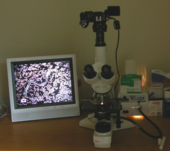
Across the ocean, Dr. Gunther Enderlein was using a microscope somewhat similar to Rife's, less complicated and less powerful. He was also able to observe live samples and to create isopathic remedies that mitigated disease conditions. These were produced by Sanum and now by a host of look alike companies. There are a few Enderlein-trained practitioners in North America, but very few and their work is now threatened by overzealous federal agents who seem to be doing a better job looking after industry than the public. A bit to the north and later, Gaston Naessens modified an Olympus microscope so he could view live blood. This resulted in yet different strategies for eliminating the causes of diseases. He has also been persecuted but he is still alive and still teaching a few students. Christopher Bird wrote a book about Naessens and referred to him as the Galileo of the microscope. So what exactly is so dangerous about looking through a microscope that people have been raided, had their equipment confiscated or destroyed, and even dragged from their beds in the middle of the night and jailed?
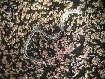
Those using darkfield microscopes may not be widely understood by their peers, but in Europe, thank goodness, after 20-25 years of education, doctors are regarded as sufficiently intelligent, responsible, and serious about their work that they can decide for themselves whether or not such instruments enhance their understanding of their patients' needs. After all, the microscope is not a treatment device. It is merely an adjunctive tool for viewing live samples and assessing the efficacy of the treatments. For instance, if a patient has spirochetes in the blood or saliva, a sample can be put on a piece of glass and viewed in such a manner as to determine whether or not the condition is resolving and by how much. I owe my life to the fact that after a very dangerous encounter with a spider, I was able to see my blood in someone's scope. If the information had not been made available, I would not have understood how to recover from the venom and bacterial neurotoxin.
Obviously, when a treatment is driven underground, it becomes harder and harder to find qualified practitioners. Those who are qualified usually have to go abroad for training and they generally work without the benefits of grants and other perks that make it possible for people within the system to survive. I believe the current abuse of authority is more than senseless. It presupposes that everyone possessed of a tool or piece of understanding that is outside one's own area of familiarity is ipso facto dangerous and deluded. This serves only to protect the vested interests of those who are profiting from the status quo. It does not support new understanding and it does not protect the public. This said, misunderstanding about the scope is rampant so a little more explanation might be helpful. The Scope There are many kinds of microscopes, some of them enlarge very little more than a magnifying glass. However, once one gets to the level of clinically useful scopes, there are really two main types of scopes: brightfield and darkfield. The way these microscopes are built is fairly straightforward. There is a light source under the slide and viewing optics over the slide. A simple scope has one objective but there are scopes with turrets holding 4, 5, and 6 objectives. These are usually of different magnifications such as 2x, 4x, or 10x for rapid screening of sample, then an objective with a little more magnification for zeroing in on the interesting parts. This might be 10x or 25x. Then, there is another more powerful objective, 40-65x, and finally usually an even more powerful objective, 100x. Just as with binoculars, cameras, and telescopes, the optic quality varies enormously from one objective to the next, but the principles are similar for all. The objective is used to magnify the object being viewed. What is different about a microscope from a magnifying glass or telescope is that there is a condenser under the sample that passes the light through the sample. In brightfield, the light is so bright that it washes out faint and transparent objects, and, in the case of blood, this has given rise to the erroneous belief that blood is sterile and that proper management of our blood supply is not necessary. However, every sensible person knows that blood is not actually sterile. If it were, there would be no reason for performing blood tests to determine the presence or absence of infections. In darkfield, the condenser splits the light so that it comes in from the edges and silouhettes the objects on the slide, thus enabling one to see what is washed out in brightfield. In reality, this is the only difference between a brightfield and darkfield microscope. The rest of the differences depend on the observer and the use the observer makes of the observations. Restricting use of a condenser that splits light is, as Christopher Bird suggested, about as reasonable as telling Galileo that his observations were making the church and state uncomfortable so he should be excommunicated for heresies and spend the rest of his life under house arrest. Surely, we are not going to repeat the mistakes of the Inquisition and brand microscopists as quacks simply because their observations are making certain people uneasy?
Monomorphism, Pleomorphism, and the New Biology One of the academic points that sometimes separates brightfield microscopists from darkfield microscopists is the issue of whether or not "germs" are pleomorphic. Banning a microscope because some people are on one side of the aisle and others on the opposite would be like saying the telescope is responsible for whether or not our solar system is geocentric or heliocentric. Obviously a piece of glass does not alter any facts about the nature of reality so the real issue is whether or not well educated and reasonable people have to agree on everything or whether some measure of exploration beyond the accepted is permissible. In our present time, getting permission is becoming increasingly difficult. To the best of my knowledge, making an observation does not constitute the practice of medicine. The practice of medicine involves the use of pharmaceutical drugs and surgery, the only domain protected by monopoly. Since doctors are not trained in acupuncture, homeopathy, nutrition, or countless other subjects, there are other specialists working in these areas whose training and qualifications are suitable to their pursuits. However, to the extent that microscopy does not involve treatment, it is not a medical device and its use cannot possibly be construed as the practice of medicine, but there are even more important reasons for preventing the demise of these microscopes, chief of which is that at this point in time, these are the only scopes that allow the user to observe live samples without the use of any dyes, fixatives, reagents, or other artifact causing substances. It would take an entire book to explain how and why other scopes contribute to certain misunderstandings because of the techniques involved in their use. For those who need to understand the arguments in greater depth, I would recommend The Living Cell by H. Hillman and P. Sartory, former council member of the Royal Microscopical Society.
Copyright by Ingrid Naiman 2008 https://www.bitchute.com/video/WyjLFvBm1bsZ/
|
||||||||||||
Home || Contact Us |
|||||||||||||
No content on any of the pages of this web site may be reproduced without written permission of Ingrid Naiman and Seventh Ray Press, publisher of this site. |
|||||||||||||
|
|||||||||||||